Home » Without Label » Left Hip Muscles Anatomy - Modified Osteotomy Of Posterolateral Overhanging Part Of The Trochanter Via Posterior Approach For Hip Arthroplasty An Anatomical Study Bmc Musculoskeletal Disorders Full Text : Small and deep muscles which mainly externally rotate the thigh at the hip joint and stabilize the pelvis.
Left Hip Muscles Anatomy - Modified Osteotomy Of Posterolateral Overhanging Part Of The Trochanter Via Posterior Approach For Hip Arthroplasty An Anatomical Study Bmc Musculoskeletal Disorders Full Text : Small and deep muscles which mainly externally rotate the thigh at the hip joint and stabilize the pelvis.
Left Hip Muscles Anatomy - Modified Osteotomy Of Posterolateral Overhanging Part Of The Trochanter Via Posterior Approach For Hip Arthroplasty An Anatomical Study Bmc Musculoskeletal Disorders Full Text : Small and deep muscles which mainly externally rotate the thigh at the hip joint and stabilize the pelvis.. They also stabilise the hip joint by 'pulling' the femoral head into the acetabulum of the pelvis. See anatomy hip muscles stock video clips. The thigh bone or femur and the pelvis join to form the hip joint. The hip's unique anatomy enables it to be both extremely strong and amazingly flexible, so it can bear weight and allow for a wide range of movement. Adductor muscles of the hip the adductor brevis, adductor longus, adductor magnus, pectineus, and gracilis make up the adductor group.
See anatomy hip muscles stock video clips. The strong muscles of the hip region also help to hold the hip joint together and prevent dislocation. The hip joint is the largest ball and socket joint in the body. He is an attending emergency medicine phys. The iliofemoral, pubofemoral, and ischiofemoral ligaments represent the thickenings of the joint capsule.
Anatomy Of Knee from marvel-b1-cdn.bc0a.com To put it plainly, sometimes hip pain comes from the hip, but a lot of times hip pain comes from the back. The iliofemoral, pubofemoral, and ischiofemoral ligaments represent the thickenings of the joint capsule. The hamstrings are three muscles at the back of the thigh that affect. There are four gluteal muscles, located at the posterior side of the hip bone: When the hip muscles are left more or less intact, they are able to support the new. The gluteus medius muscle helps abducts the thigh along with the gluteus maximus, but can rotate the thigh inward where the gluteus maximus rotates the thigh outward. In utero fetal hips lie typically in flexion, abduction and external rotation, with the left hip usually muscular anatomy. The socket in the pelvis, is called the acetabulum (figure 1.3).
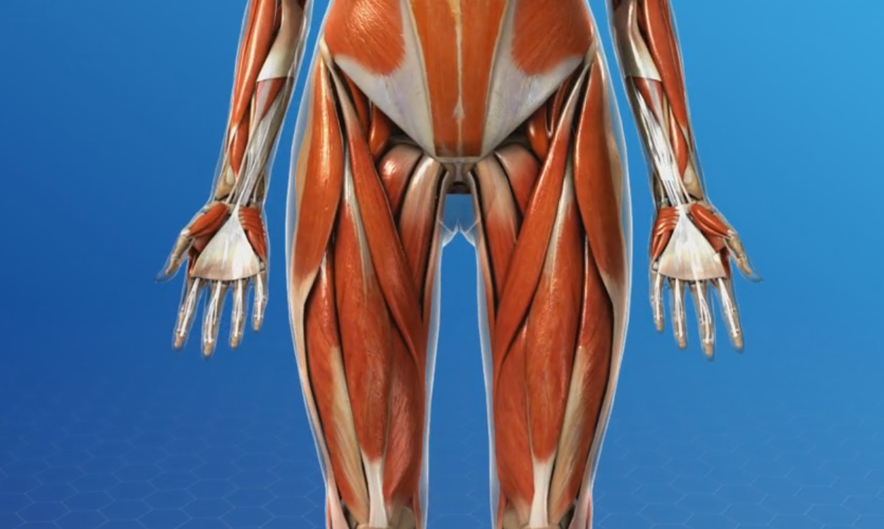
Ebraheim's educational animated video describes the muscle anatomy of the hip and buttocks region with simple images;
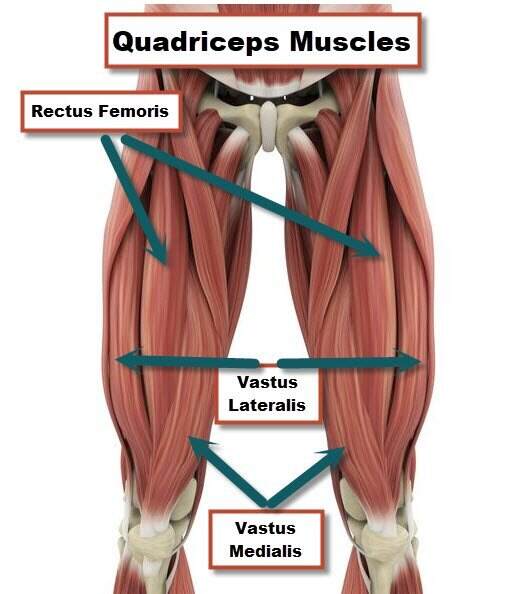
Of the quadriceps muscles, it has the least affect on flexion of the knee. The hip joint is a ball and socket synovial joint, formed by an articulation between the pelvic acetabulum and the head of the femur. These are the gluteus maximus, gluteus medius, gluteus minimus, and tensor fasciae latae. The hip flexors are several muscles that bring your legs and trunk together in a flexion movement. Anatomy it band pelvis muscle pelvis with muscles hip muscles muscles of pelvis tensor fascia latae psoas major anatomy pelvis tensor fascia lata pelvis muscles. (2017, elsevier) should be consulted. Any injury or disease of the hip will. The general action of these muscles is to laterally rotate the lower limb. Adductor muscles of the hip the adductor brevis, adductor longus, adductor magnus, pectineus, and gracilis make up the adductor group. The anatomy of the hip and back is comprised of numerous parts that can be injured or wear out, and many problems that occur in this area can display the exact same symptoms or pathology. There are also diseases and disorders that can cause the pain to. Small and deep muscles which mainly externally rotate the thigh at the hip joint and stabilize the pelvis. This blog post article is an overview of the muscles of the pelvis.
Adductor muscles of the hip the adductor brevis, adductor longus, adductor magnus, pectineus, and gracilis make up the adductor group. It's formed by the joining of three muscles: This blog post article is an overview of the muscles of the pelvis. This video also provides you with a. It is also referred to as a ball and socket joint and is surrounded by muscles, ligaments, and tendons.
What Is A Hip Flexor Plano Orthopedic Sports Medicine Center from www.posmc.com These are the gluteus maximus, gluteus medius, gluteus minimus, and tensor fasciae latae. Gently lower your left leg on the floor. The hip flexors are several muscles that bring your legs and trunk together in a flexion movement. The muscles you probably know the best are your glutes. If soft tissue, such as skin, muscles, fat, and fascia get strained or injured, left hip pain can come from the abdominal wall. You can strain or tear your hip flexor muscles through sudden movements or falls. This video also provides you with a. When the hip muscles are left more or less intact, they are able to support the new.
Left hip muscles anatomy :
The ball is the head of the femur (thigh bone). Both the ball and socket are lined with smooth cartilage which allows the bones to slide against each other easily (figure 1.3). The muscles are broken down into three layers, and are primarily used to assist with the breathing process. Gluteus maximus, gluteus medius, gluteus minimus, and tensor fasciae latae. He is an attending emergency medicine phys. It is a synergist for the gluteus medius. Ebraheim's educational animated video describes the muscle anatomy of the hip and buttocks region with simple images; This muscle attaches to the kneecap. They also stabilise the hip joint by 'pulling' the femoral head into the acetabulum of the pelvis. The below the gluteus medius are several muscles, one of which is the gluteus minimus, the smallest of the gluteal muscles. The hip joint is a ball and socket synovial joint, formed by an articulation between the pelvic acetabulum and the head of the femur. The hip joint allows for movement in three major axes, all of which are perpendicular to one another. The movements available at the hip include:
It's formed by the joining of three muscles: Gluteus maximus, gluteus medius, gluteus minimus, and tensor fasciae latae. (2017, elsevier) should be consulted. The transverse axis permits flexion and extension movement. This video also provides you with a.
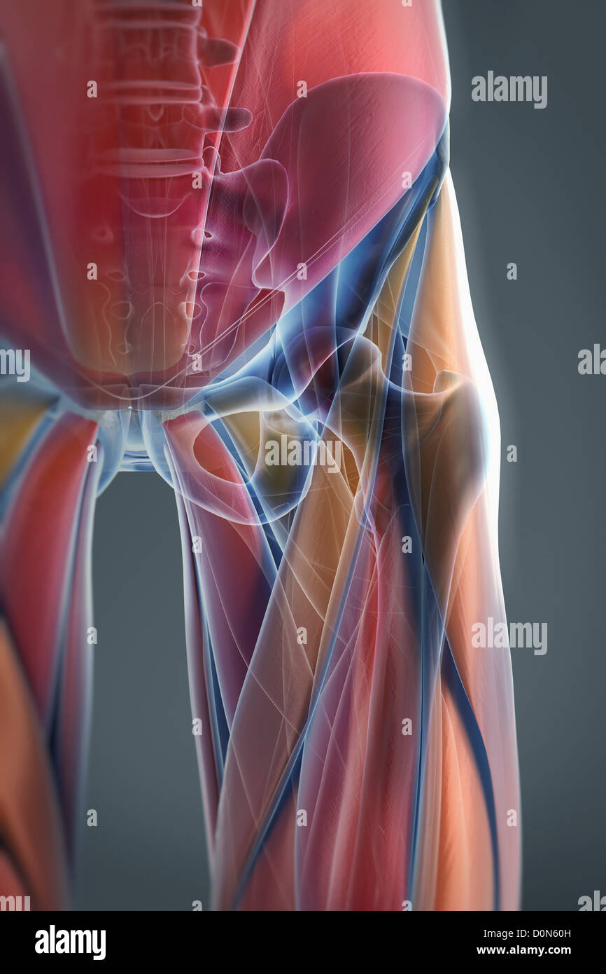
A Transparent Skin Reveals The Muscles And Skeletal Structures Of The Left Hip Joint The Bones Have An X Ray Appearance Stock Photo Alamy from c8.alamy.com The thigh bone or femur and the pelvis join to form the hip joint. This video also provides you with a. The muscles are broken down into three layers, and are primarily used to assist with the breathing process. When the hip muscles are left more or less intact, they are able to support the new. There are four gluteal muscles, located at the posterior side of the hip bone: Ligaments, tendons, and muscles play an important role in the function of the hip. The location of the center of the entire axis is at the femoral head. Ebraheim's educational animated video describes the muscle anatomy of the hip and buttocks region with simple images;
Any injury or disease of the hip will.
The strong muscles of the hip region also help to hold the hip joint together and prevent dislocation. It is also referred to as a ball and socket joint and is surrounded by muscles, ligaments, and tendons. The gluteal muscles consist of the gluteus maximum, gluteus medius, and gluteus minimus. The iliofemoral, pubofemoral, and ischiofemoral ligaments represent the thickenings of the joint capsule. The hip flexors are several muscles that bring your legs and trunk together in a flexion movement. (2017, elsevier) should be consulted. There are also diseases and disorders that can cause the pain to. The quadriceps muscles are four powerful muscles at the front of the thigh involved in movement. These are the gluteus maximus, gluteus medius, gluteus minimus, and tensor fasciae latae. It is a synergist for the gluteus medius. If soft tissue, such as skin, muscles, fat, and fascia get strained or injured, left hip pain can come from the abdominal wall. Gently lower your left leg on the floor. They allow you to move your leg or knee up towards your torso, as well as to bend your torso forward at the hip.